

The best ophthalmic equipment and instruments in 2026 cover visual acuity, refraction, anterior-eye examination, pressure measurement, and retinal imaging. For most clinics, a slit lamp, autorefractor, phoropter, tonometer, and acuity chart form the core. OCT and fundus imaging belong where patient mix and referrals justify them.
“Best” means clinically appropriate, repeatable, serviceable, and workable during a Tuesday. This ranking weighs use, training, maintenance, and ownership cost. It covers ten types, not versions of one machine.
WHO reports 2.2 billion people have vision impairment; 1 billion cases remain unaddressed or preventable (World Health Organization, February 10, 2026). Annual productivity losses reach US$411 billion.
| Rank | Equipment | Primary role | Check first |
|---|---|---|---|
| 1 | Slit lamp | Anterior examination | Optics, ergonomics |
| 2 | Autorefractor/keratometer | Objective refraction | Repeatability |
| 3 | Tonometer | IOP measurement | Calibration |
| 4 | OCT | Retinal imaging | Software, service |
| 5 | Fundus camera | Retinal records | Image gradability |
| 6 | Phoropter | Subjective refraction | Connectivity |
| 7 | Lensmeter | Lens verification | Progressive handling |
| 8 | Ophthalmoscope/retinoscope | Portable checks | Beam quality |
| 9 | Acuity system | Vision testing | Room distance |
| 10 | Ophthalmic unit | Positioning | Load, travel |
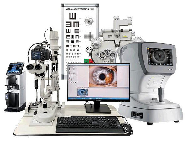
The slit lamp is the examination-room workhorse for anterior structures. LINK INSTRUMENTS offers slit lamp microscopes, including the SL-5A with five-step magnification and warm LED illumination. Buy digital capture only when documentation or teaching will use it.
2. Autorefractor/Keratometer
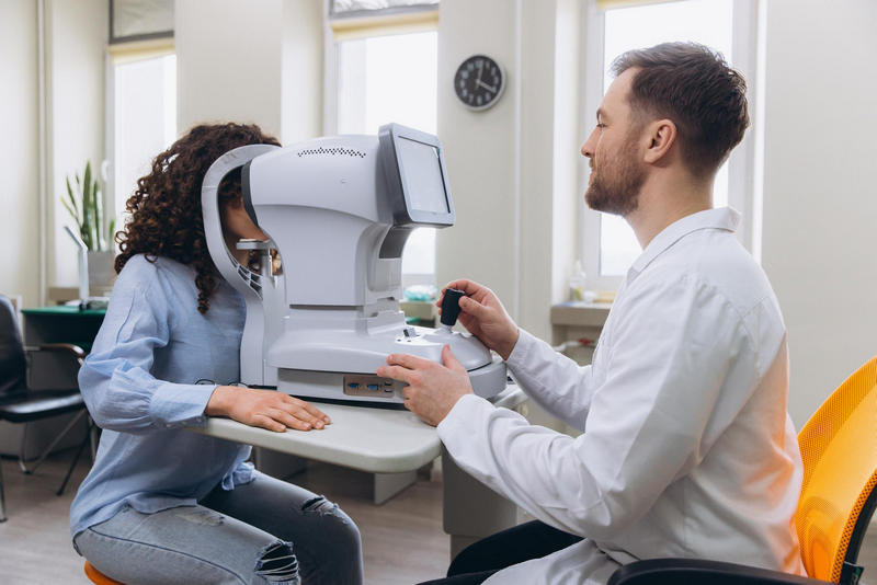
An autorefractor supplies an objective starting point; keratometry adds corneal curvature. It does not replace subjective refinement. The LINK ARK-1800 portable autorefractor pairs a seven-inch display with a thermal printer. Test repeatability on real patients, including small pupils and high ametropia.
Every general clinic needs intraocular-pressure measurement. Non-contact models suit screening; rebound instruments aid mobility; Goldmann applanation remains a reference. Contact applanation is “most accurate, typically a Goldmann tonometer,” says the American Academy of Ophthalmology, February 11, 2026. Check calibration before comparing LINK tonometer options.
OCT provides cross-sectional images of the retina, nerve fibre layer, and optic nerve head. It earns its space in glaucoma and medical-retina practices, but not in a refraction-only shop. Compare segmentation, normative databases, exports, licences, and engineer response—not scan speed alone. LINK lists the OCT-500 platform.
A fundus camera records the retina and optic disc for comparison and screening. The LINK HFC-200 handheld fundus camera lists a 45-degree field. Ask for unedited images from the population you serve; a handsome camera with poorly gradable output is expensive furniture.
The phoropter turns objective findings into a prescription through subjective comparison. Manual models are economical and easy to troubleshoot. Digital units speed lens changes and data transfer when devices communicate properly. LINK’s DVT-1000 auto phoropter supports LCD charts and tablet control. Keep trial lenses as backup.
A lensmeter verifies sphere, cylinder, axis, prism, and addition before dispensing. Progressive lenses can expose weak detection quickly. The LINK AL-860 automatic lensmeter uses a green LED and automatic lens identification. Test your real lens mix, not one demonstration lens.
These small instruments still earn their place. A direct ophthalmoscope supports quick disc checks; an indirect model gives a wider retinal view. A retinoscope helps when patient responses are unreliable, especially in paediatrics. LINK supplies wall-mounted and handheld diagnostic sets. Judge beam clarity, charging, and balance.
Visual acuity starts almost every examination. LCD charts save space; projectors work when distance and ambient light are controlled. Match local notation, language, paediatric needs, and room geometry. LINK’s visual acuity systems include LCD, projected, and near-vision options. Measure installation distance—do not guess.
Support equipment gets little glory, but poor positioning slows every test. A stable unit should suit patient height, clinician reach, instrument weight, and wheelchair access. The LINK CT-1000 ophthalmic unit combines electric chair movement, tabletop travel, and a phoropter arm. Confirm load, voltage, footprint, and service access.
Start with patient volume and services, then build around the exam sequence.
Map the pathway: List each test, operator, handoff, and referral.
Verify compliance: Request certificates for the exact model and destination.
Run a trial: Check repeatability, positioning, cleaning, export, and restart time.
Price five years: Include software, calibration, consumables, training, parts, and downtime.
Audit support: Get contacts, response times, manuals, and parts availability in writing.
The Ophthalmology Foundation’s January 30, 2026 guidance calls slit lamps and tonometers common equipment, while OCT and fundus cameras are specialized. Buy daily essentials first, demand model-level compliance documents, and conduct acceptance testing after installation.
Buy an acuity system, slit lamp, autorefractor, phoropter or trial lenses, and tonometer first. Add retinal imaging when caseload supports it.
No. OCT suits retinal, glaucoma, and monitoring workflows. A basic optometry room may refer these cases.
Follow manufacturer instructions and local policy. Abnormal drift or impact requires immediate assessment.
Choose manual equipment for simple maintenance. Choose digital when patient volume and integration justify it.
Review the LINK INSTRUMENTS ophthalmic equipment range, then request current specifications, certificates, warranty terms, training, and a destination-specific quotation.
For most general clinics, the best choice is a slit lamp, autorefractor/keratometer, tonometer, phoropter, and acuity display. Add a fundus camera when screening is the gap; choose OCT when glaucoma or retinal disease drives demand. LINK INSTRUMENTS merits a shortlist where OEM/ODM matters, but the winner must pass a clinical trial, compliance review, and five-year cost check.